FUBP1
FUBP1,全稱為Far upstream element-binding protein 1,是一種單鏈DNA結合蛋白,常見別名包括FUSE結合蛋白1。該蛋白能夠結合到DNA上的特定序列,調節基因的表達,參與細胞增殖、分化和凋亡等過程。FUBP1在多種癌癥中都有異常表達,與腫瘤的發生和發展密切相關。研究表明,FUBP1與FXR1蛋白相互作用,影響FUBP1的表達和活性,進而影響腫瘤細胞的增殖、遷移和侵襲,并降低其對化療藥物的敏感性。FUBP1的表達水平也與多種疾病的發生發展有關,如肺鱗狀細胞癌、腎透明細胞癌和乳腺癌等。針對FUBP1的藥物研發正在積極推進中,例如FUBP1-IN-1是一種有效的FUBP1抑制劑,能夠干擾FUBP1與單鏈目標DNA的FUSE序列結合,展現出在腫瘤治療中的潛力。此外,FUBP1的表達水平也可作為生物標志物,用于預測患者對化療的反應,為臨床治療提供參考。
熱銷產品
FUBP1 Recombinant Monoclonal Antibody (CSB-RA157765A0HU)
驗證數據
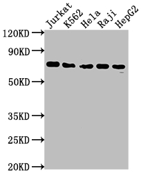
Western Blot
Positive WB detected in: Jurkat whole cell lysate, K562 whole cell lysate, Hela whole cell lysate, Raji whole cell lysate, HepG2 whole cell lysate
All lanes: FUBP1 antibody at 1:2000
Secondary
Goat polyclonal to rabbit IgG at 1/50000 dilution
Predicted band size: 68, 69 kDa
Observed band size: 69 kDa
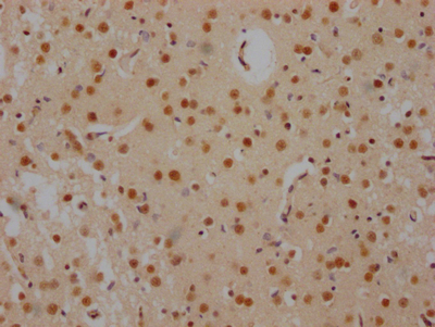
IHC image of CSB-RA157765A0HU diluted at 1:100 and staining in paraffin-embedded human brain tissue performed on a Leica BondTM system. After dewaxing and hydration, antigen retrieval was mediated by high pressure in a citrate buffer (pH 6.0). Section was blocked with 10% normal goat serum 30min at RT. Then primary antibody (1% BSA) was incubated at 4℃ overnight. The primary is detected by a Goat anti-rabbit IgG polymer labeled by HRP and visualized using 0.05% DAB.
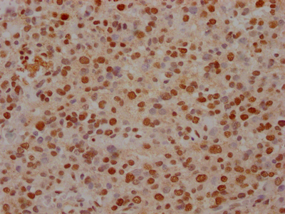
IHC image of CSB-RA157765A0HU diluted at 1:100 and staining in paraffin-embedded human glioma cancer performed on a Leica BondTM system. After dewaxing and hydration, antigen retrieval was mediated by high pressure in a citrate buffer (pH 6.0). Section was blocked with 10% normal goat serum 30min at RT. Then primary antibody (1% BSA) was incubated at 4℃ overnight. The primary is detected by a Goat anti-rabbit IgG polymer labeled by HRP and visualized using 0.05% DAB.
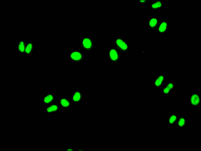
Immunofluorescence staining of Hela Cells with CSB-RA157765A0HU at 1:50, counter-stained with DAPI. The cells were fixed in 4% formaldehyde, permeated by 0.2% TritonX-100, and blocked in 10% normal Goat Serum. The cells were then incubated with the antibody overnight at 4℃. Nuclear DNA was labeled in blue with DAPI. The secondary antibody was FITC-conjugated AffiniPure Goat Anti-Rabbit IgG (H+L).
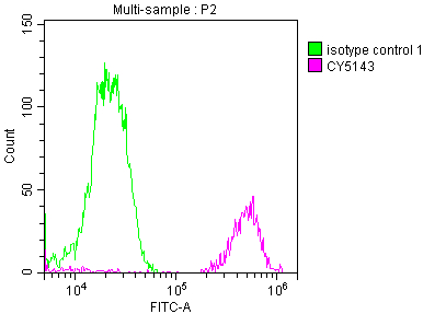
Overlay histogram showing Jurkat cells stained with CSB-RA157765A0HU (red line) at 1:50. The cells were fixed with 70% Ethylalcohol (18h) and then incubated in 10% normal goat serum to block non-specific protein-protein interactions followedby the antibody (1μg/1*106 cells) for 1 h at 4℃.The secondary antibody used was FITC-conjugated goat anti-rabbit IgG (H+L) at 1/200 dilution for 30min at 4℃. Control antibody (green line) was Rabbit IgG (1μg/1*106 cells) used under the same conditions. Acquisition of >10,000 events was performed.
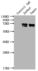
Immunoprecipitating FUBP1 in Jurkat whole cell lysate
Lane 1: Rabbit control IgG instead of CSB-RA157765A0HU in Jurkat whole cell lysate.
For western blotting,a HRP-conjugated Protein G antibody was used as the secondary antibody (1/2000)
Lane 2: CSB-RA157765A0HU(2μg)+ Jurkat whole cell lysate(500μg)
Lane 3: Jurkat whole cell lysate (10μg)
FUBP1 Antibodies
FUBP1 for Homo sapiens (Human)
| 產品貨號 | 產品名稱 | 種屬反應性 | 應用類型 |
|---|---|---|---|
| CSB-RA157765A0HU | FUBP1 Recombinant Monoclonal Antibody | Human | ELISA, WB, IHC, IF, FC, IP |
| CSB-RA094548A0HU | FUBP1 Recombinant Monoclonal Antibody | Human, Mouse, Rat | ELISA, WB, FC, ICC |
FUBP1 Proteins
FUBP1 Proteins for Homo sapiens (Human)
| 產品貨號 | 產品名稱 | 來源 |
|---|---|---|
| CSB-YP846584HU CSB-EP846584HU CSB-BP846584HU CSB-MP846584HU CSB-EP846584HU-B |
Recombinant Human Far upstream element-binding protein 1 (FUBP1) | Yeast E.coli Baculovirus Mammalian cell In Vivo Biotinylation in E.coli |