SMAD1
SMAD1是SMAD蛋白家族的成員之一,全稱為SMAD家族成員1,也常被稱為MADH1或hMAD-1。它在細胞內承擔著信號傳遞的關鍵角色:當骨形態(tài)發(fā)生蛋白(BMP)等外部信號與細胞膜受體結合后,SMAD1會被激活并進入細胞核,參與調控一系列基因的表達,進而影響細胞的生長、分化、凋亡,以及組織器官的發(fā)育,尤其在骨骼形成和胚胎早期發(fā)育中作用顯著。
若SMAD1功能異常,可能引發(fā)多種疾病。比如先天性骨骼發(fā)育缺陷,因其在骨形成中的核心作用;在肺癌、乳腺癌等癌癥中,其表達變化常與腫瘤細胞增殖、轉移能力增強相關;此外,它還參與血管內皮細胞功能調節(jié),異常時可能影響血管健康。目前針對SMAD1所在通路的藥物研發(fā)處于活躍階段,一些小分子抑制劑或靶向抗體正處于臨床前或早期臨床研究,通過調節(jié)通路活性間接影響SMAD1功能,有望為相關疾病治療提供新方向。
熱銷產品
SMAD1 Recombinant Monoclonal Antibody (CSB-RA207404A0HU)
驗證數據
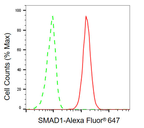
Flow cytometric analysis of SMAD1 expression in HAP-1 cells using SMAD1 antibody. Green, isotype control; red, SMAD1.
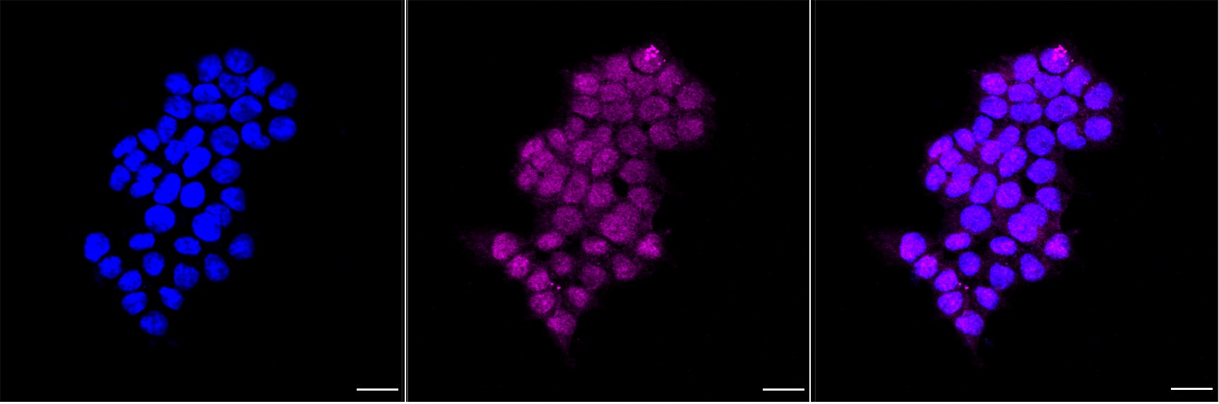
Immunocytochemical staining of HAP-1 cells with SMAD1 antibody. Nuclei were stained blue with DAPI; SMAD1 was stained magenta with Alexa Fluor? 647. Images were taken using Leica stellaris 5. Protein abundance based on laser Intensity and smart gain: Medium. Scale bar, 20 μm.
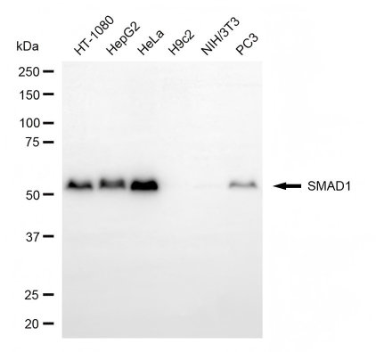
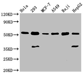
Western blotting analysis using SMAD1 antibody. Total cell lysates (30 μg) from various cell lines were loaded and separated by SDS-PAGE. The blot was incubated with SMAD1 antibody and HRP-conjugated goat anti-rabbit secondary antibody respectively.
SMAD1 Antibody (CSB-PA618998LA01HU)
驗證數據
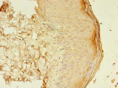
Immunohistochemistry of paraffin-embedded human skin tissue using CSB-PA618998LA01HU at dilution of 1:100
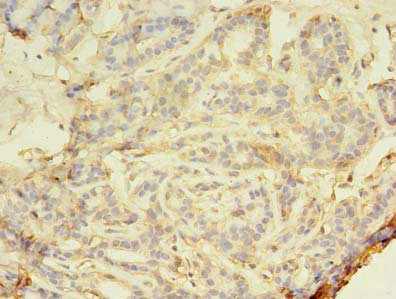
Immunohistochemistry of paraffin-embedded human breast cancer using CSB-PA618998LA01HU at dilution of 1:100
Western Blot
Positive WB detected in: Hela whole cell lysate, 293 whole cell lysate, MCF-7 whole cell lysate, A549 whole cell lysate, Raji whole cell lysate, HepG2 whole cell lysate
All lanes: SMAD1 antibody at 3µg/ml
Secondary
Goat polyclonal to rabbit IgG at 1/50000 dilution
Predicted band size: 53, 16 kDa
Observed band size: 53 kDa
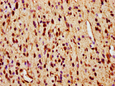
IHC image of CSB-PA618998LA01HU diluted at 1:600 and staining in paraffin-embedded human glioma performed on a Leica BondTM system. After dewaxing and hydration, antigen retrieval was mediated by high pressure in a citrate buffer (pH 6.0). Section was blocked with 10% normal goat serum 30min at RT. Then primary antibody (1% BSA) was incubated at 4°C overnight. The primary is detected by a biotinylated secondary antibody and visualized using an HRP conjugated SP system.
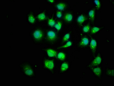
Immunofluorescence staining of Hela cells with CSB-PA618998LA01HU at 1:200, counter-stained with DAPI. The cells were fixed in 4% formaldehyde, permeabilized using 0.2% Triton X-100 and blocked in 10% normal Goat Serum. The cells were then incubated with the antibody overnight at 4°C. The secondary antibody was Alexa Fluor 488-congugated AffiniPure Goat Anti-Rabbit IgG(H+L).
SMAD1 Antibodies
SMAD1 for Homo sapiens (Human)
| 產品貨號 | 產品名稱 | 種屬反應性 | 應用類型 |
|---|---|---|---|
| CSB-PA021786GA01HU | SMAD1 Antibody | Human,Mouse,Rat | ELISA,WB |
| CSB-PA050193 | Phospho-SMAD1 (Ser465) Antibody | Human,Mouse,Rat | ELISA,WB |
| CSB-PA135678 | SMAD1 (Ab-465) Antibody | Human,Mouse,Rat | ELISA,WB,IF |
| CSB-PA224827 | Phospho-SMAD1 (Ser206) Antibody | Human,Mouse,Rat | ELISA,WB |
| CSB-PA056920 | Phospho-SMAD1 (Ser187) Antibody | Human,Mouse | ELISA,WB |
| CSB-PA927298 | SMAD1 (Ab-187) Antibody | Human,Mouse | ELISA,WB,IHC,IF |
| CSB-PA998747 | SMAD1 Antibody | Human,Mouse,Rat | ELISA,WB |
| CSB-PA000674 | Phospho-SMAD1 (S465) Antibody | Human | IHC, ELISA |
| CSB-PA004104 | SMAD1 Antibody | Human,Mouse | WB, IHC, IF, ELISA |
| CSB-PA005538 | SMAD1 Antibody | Human,Mouse,Rat | WB, ELISA |
| CSB-PA009920 | Phospho-SMAD1 (S187) Antibody | Human,Mouse,Monkey | WB, IHC, ELISA |
| CSB-PA009928 | SMAD1 Antibody | Human,Mouse,Rat,Monkey | WB, IHC, ELISA |
| CSB-PA042170 | SMAD1 Antibody | Human,Mouse,Rat | ELISA,IHC |
| CSB-PA841004 | SMAD1 Antibody | Human,Mouse,Rat | ELISA,WB,IHC |
| CSB-PA618998LA01HU | SMAD1 Antibody | Human | ELISA, WB, IHC, IF |
| CSB-PA618998LD01HU | SMAD1 Antibody, Biotin conjugated | Human | ELISA |
| CSB-PA618998LC01HU | SMAD1 Antibody, FITC conjugated | Human | |
| CSB-PA618998LB01HU | SMAD1 Antibody, HRP conjugated | Human | ELISA |
| CSB-PA618998HA01HU | SMAD1 Antibody | Human | ELISA, IHC, IF |
| CSB-PA618998HB01HU | SMAD1 Antibody, HRP conjugated | Human | ELISA |
| CSB-PA618998HC01HU | SMAD1 Antibody, FITC conjugated | Human | |
| CSB-RA056935A0HU | SMAD1 Recombinant Monoclonal Antibody | Human | ELISA, WB, IF, FC |
| CSB-RA207404A0HU | SMAD1 Recombinant Monoclonal Antibody | Human | ELISA, WB, FC, ICC |
SMAD1 Proteins
SMAD1 Proteins for Mus musculus (Mouse)
| 產品貨號 | 產品名稱 | 來源 |
|---|---|---|
| CSB-YP021786MO CSB-EP021786MO CSB-BP021786MO CSB-MP021786MO CSB-EP021786MO-B |
Recombinant Mouse Mothers against decapentaplegic homolog 1 (Smad1) | Yeast E.coli Baculovirus Mammalian cell In Vivo Biotinylation in E.coli |
SMAD1 Proteins for Rattus norvegicus (Rat)
| 產品貨號 | 產品名稱 | 來源 |
|---|---|---|
| CSB-YP021786RA CSB-EP021786RA CSB-BP021786RA CSB-MP021786RA CSB-EP021786RA-B |
Recombinant Rat Mothers against decapentaplegic homolog 1 (Smad1) | Yeast E.coli Baculovirus Mammalian cell In Vivo Biotinylation in E.coli |
SMAD1 Proteins for Homo sapiens (Human)
| 產品貨號 | 產品名稱 | 來源 |
|---|---|---|
| CSB-YP618998HU CSB-EP618998HU CSB-BP618998HU CSB-MP618998HU CSB-EP618998HU-B |
Recombinant Human Mothers against decapentaplegic homolog 1 (SMAD1) | Yeast E.coli Baculovirus Mammalian cell In Vivo Biotinylation in E.coli |
SMAD1 Proteins for Bos taurus (Bovine)
| 產品貨號 | 產品名稱 | 來源 |
|---|---|---|
| CSB-YP628618BO CSB-EP628618BO CSB-BP628618BO CSB-MP628618BO CSB-EP628618BO-B |
Recombinant Bovine Mothers against decapentaplegic homolog 1 (SMAD1) | Yeast E.coli Baculovirus Mammalian cell In Vivo Biotinylation in E.coli |
SMAD1 Proteins for Danio rerio (Zebrafish) (Brachydanio rerio)
| 產品貨號 | 產品名稱 | 來源 |
|---|---|---|
| CSB-YP862301DIL CSB-EP862301DIL CSB-BP862301DIL CSB-MP862301DIL CSB-EP862301DIL-B |
Recombinant Danio rerio Mothers against decapentaplegic homolog 1 (smad1) | Yeast E.coli Baculovirus Mammalian cell In Vivo Biotinylation in E.coli |
SMAD1 Proteins for Coturnix coturnix japonica (Japanese quail) (Coturnix japonica)
| 產品貨號 | 產品名稱 | 來源 |
|---|---|---|
| CSB-YP872775DXJ CSB-EP872775DXJ CSB-BP872775DXJ CSB-MP872775DXJ CSB-EP872775DXJ-B |
Recombinant Coturnix coturnix japonica Mothers against decapentaplegic homolog 1 (SMAD1) | Yeast E.coli Baculovirus Mammalian cell In Vivo Biotinylation in E.coli |